Sleep Heart Health Study
6.6.2.3 EEG arousal
Scoring arousals is based on “A Preliminary Report from the Sleep Disorders Atlas Task Force of the American Sleep Disorders Association” Sleep, vol. 15, no 2, 1992. (ASDA criteria)
The scoring of EEG arousals is independent from the scoring of sleep stages (i.e. an arousal can be scored in an epoch of recording which would be classified as wake by R & K criteria). An arousal can proceed to the wake stage (by R & K criteria) or can be followed by a return to sleep.
Definition of Arousal:
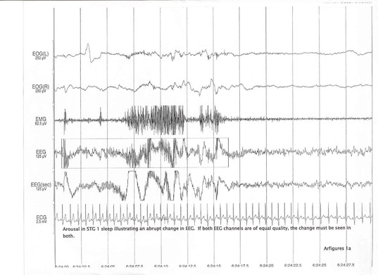
An EEG arousal is an abrupt shift in EEG frequency, which may include alpha and/or theta waves and/or delta waves and/or frequencies greater than 16 Hz lasting at least 3 s., and starting after at least 10 continuous seconds of sleep. (ArFigures 1a and 1b)
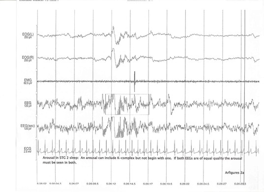
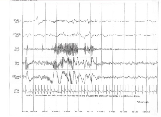
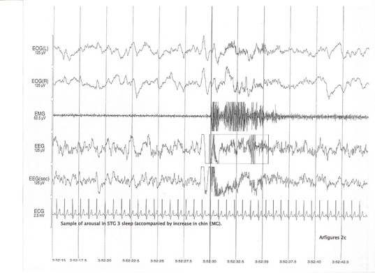
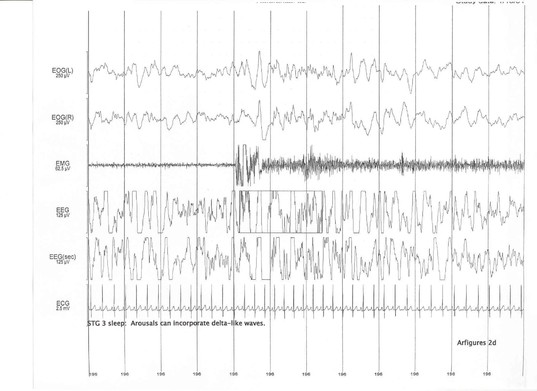
Artifacts, K complexes and delta waves are included in meeting the 3 s. duration criteria only when they occur within the EEG frequency shift (change in frequency must be visible before these waveforms). A "K" complex or spindle occurring immediately prior to the EEG shift or following is not included in the arousal duration. (ArFigures 2a, 2b, 2c and 2d)
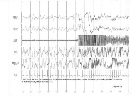
Parts of the EEG totally obscured by EMG artifact are considered an arousal if the change in background EEG in addition to the area obscured by EMG is at least > 3 sec. (ArFigure 2e)
Alpha activity of less than 3 s. duration in Non-REM sleep at a rate greater than one burst per 10 s. is not scored as an EEG arousal. Three seconds of alpha sleep is not scored as an arousal unless a 10 s. episode of alpha free sleep precedes this.
If both central EEGs are equivalent and interpretable, the arousal (EEG frequency shift) must be observed in both channels. If observed in only one of two equivalent channels, the change in EEG is assumed to be artifact and not an arousal.
Arousals lasting > 15 s. and containing awake EEG within an epoch cause the epoch to be classified as AWAKE.
TIPS for Arousals Generally:
- When unsure if change in background EEG represents an abrupt change, look at a 60 sec epoch and note if there is a discrete change from background EEG.
- Note whether changes were evident on both EEG channels.
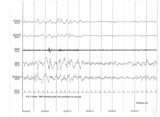
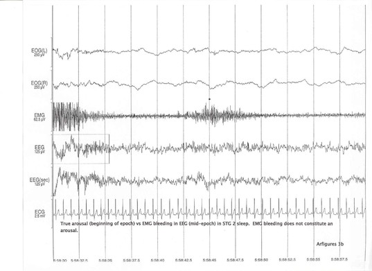
- Be careful to distinguish an increase in EEG frequency from EMG artifact (esp. in delta sleep). (ArFigures 3a and 3b).
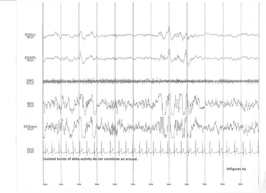
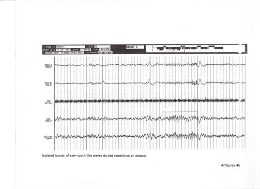
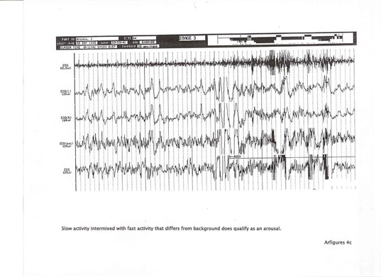
- Isolated bursts of delta activity or sawtooth-like waves do not constitute an arousal. In contrast, slow waves intermixed with fast activity that differs from background do qualify as arousals. (ArFigures 4a, 4b and 4c.)
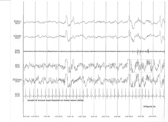
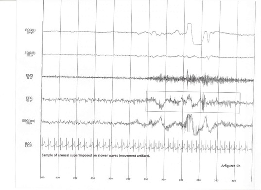
- Occasionally, EEG acceleration is superimposed on slower waves. The slowing may be an artifact secondary to movement or burst of delta waves. If there is evidence of embedded EEG acceleration for > 3 sec., mark as an arousal. (ArFigures 5a and 5b)
Arousals in REM
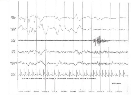
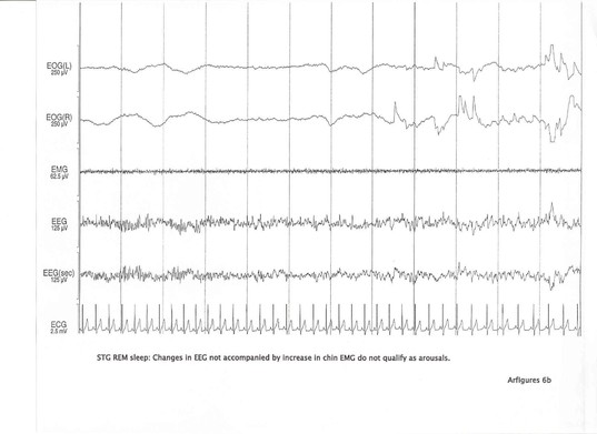
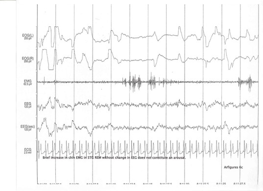
In stage REM, an EEG frequency shift must be accompanied by a simultaneous increase in amplitude of the chin EMG (lasting over 0.5 s.). An arousal starts when a definite change in background EEG is visualized. The increase in the chin EMG can occur anytime during the arousal (can be at the end) and is not a marker for the beginning of the arousal. However, increased EMG activity without a change in background EEG does not constitute an arousal (ArFigures 6a, 6b and 6c).
TIPS for Arousals in REM:
- If the level of REM EMG appears to be fluctuating, then the increase in EMG in the area of a putative arousal needs to be more than the background level of fluctuations, to identify this as a REM arousal.
- A long period of alpha activity before an EMG increase may mark the beginning of the arousal if the alpha activity represents the change in the background pattern.
Potential Problems:
Some studies with high RDIs have many arousals that may appear to last > 15 sec. within given epochs. If all such epochs were classified as AWAKE, then the respiratory events would not be included in the RDI and the RDI will be underestimated. When faced with this situation, the scorer may attempt to keep the duration of the arousal to as short as feasible (e.g., corresponding to the length of the waking EEG in that epoch). This will maximize the number of epochs containing arousals that are captured as "sleep".
